|
Health Consequences of D. U. weapons used by U.S.
and British Forces
Abstract:
A case control study of cancer cases in military
troops and its relation to Depleted Uranium used by U.S. and British
forces.
The study was a statistically and clinically descriptive
study of cancer cases in Iraq. These studies involved Iraqi military
personnel in the southern region of Iraq following the Gulf War.
1425 cases were selected retrospectively, which were
diagnosed with different types of cancer in military and civilian
hospitals. The study included cases of males who had participated
in the war in the southern region of Iraq. The cases included
only males between the ages of 19 and 50.
The study includes tabulation of cancer cases in
this age group to see whether there is a positive association
or odds ratio between those cases and D.U. used by the U.S. and
British forces.
It is estimated that about 630,000 pounds of depleted
uranium contaminated huge areas including targets and the surrounding
environment. The study shows increasing registration of different types of cancer cases and change in the epidemiological pattern of there occurrence with time among military personnel who were in the southern region of Iraq during the war. There is a significant correlation and association between these cases and D.U. exposure. This is illustrated by odds ratio of lymphomas and leukemias were 5.6 and 4.8 respectively.
Introduction:
In the war against Iraq in 1991 allied forces used
630,000 pounds of depleted uranium. Consequently, there are adverse
health effects on human beings and the environment in Iraq and
probably in the neighboring countries such as Kuwait and Saudi
Arabia.
Military personnel and their families were exposed
to depleted uranium weapons, which have a radiological and chemically
toxic effect secondary to the their explosions. In addition,
it is a source of contamination of the environment.
The central committee that was established in Iraq
did a series of epidemiological investigations and field attachment
surveys on the ill effects of depleted uranium weapons on human
beings and the environment.
The result of this investigation is that both military
personnel and their families who have been exposed to depleted
uranium have experienced radiological and chemically toxic effects.
Health problems may also exist in Kuwait, as a result of the
huge contaminated area. It is estimated that approximately 14
acres, which are being used as a collection point of contaminated
and destroyed vehicles and tanks. In addition, huge unknown quantities
of D.U. munitions were buried in the ground in this area. (Dan
Fahey, 1998).
There is also a probability of pollution in Saudi
Arabia. Under the command of the New Jersey Company of American
National Army, King Khalid Military City and King Abdul Aziz Air
Force Base were used as collecting points for contaminated and
destroyed vehicles and tanks.
Objectives:
Material and Method: A case control study of different types of cancer cases observed in military troops who participated in the war in the southern region of Iraq in 1991. To determine relative risk (odds ratio) of these cases, military personnel with cancer was compared to cases free from cancer in order to study the factor incriminated. The sample of cancer cases selected includes lymphomas, leukemias, lung CA, bone CA, gastrointestinal cancers, brain CA, and liver CA. The cases studied are those which are registered retrospectively from military and civilian hospitals in the age group (19-50 years) who were in service at that time. The cases were identified clearly from the case sheets and the addresses of their residence which were fixed since 1991 up to 1997. 1425 people, whose residences were known, were asked if they were exposed to an explosion. Sample comparative groups were identified and selected from military patients free of cancer who were admitted to the military hospitals from the same age group and sex. Their addresses were fixed from the case sheets and they were asked whether they were exposed to an explosion while in field operation. Method of selection of sample cases was done through study of all cancer cases that were diagnosed and registered from 1991-1997. Size of sample: It includes all recorded cancer cases that were diagnosed and registered in the civilian and military hospitals. Data collection: It included case history, clinical exam, laboratory investigation, radiological finding and results of histopathological findings through sample form attached in the appendix A. Direct interviews were done with control group. Each were asked about exposure to explosion. Analysis of sample forms was done manually. Presentation of data in form of tables and histograms.
Results:
Table (1):
Different types of cancer cases that were registered
from 1991 through 1997 of military participants exposed to depleted
uranium.
Shows an increase in registration of different types of cancer
cases from 1991 through 1997 in those who were exposed to explosions
in the battle field including lymphomas, leukemias, lung cancer,
and brain cancer.
Table (2):
Different cancer cases that were registered from 1991 through
1997 in military participants not exposed to the causative agent
under study (D.U.)
Shows an increase in registration of different types of cancer
cases starting in the year 1992 for those military personnel who
were not exposed to an explosion in the field in comparison with
1991, especially in relation to lung cancer, gastrointestinal
cancer, leukemias, and lymphomas.
Table (3):
Relative Risk is estimated by calculating odds ratio of different
types of cancer cases.
Shows the odds ratio which is "the degree of association between the factor under study and cancer cases."
Statistically, it is significant in relation to lymphomas, leukemia,
lung , brain, gastrointestinal, bone, and liver cancers.


Discussion: The tables and graphs indicate there is change in the pattern of different types of cancer cases in military personnel who were involved in the southern region of Iraq during the war with U.S. and British
forces in 1991.
The sample cases were from military personnel who
participated and were exposed to explosions of D.U. weapon in
the south of Iraq, Rumaila north, Rumaila south, strategic pipeline
near pump station (Border of Saudi Arabia), and the road between
Safwan and Kuwait at which attacks on tanks and vehicles continued
2 days after withdrawal of Iraqi troops.
U.S. and British forces dropped about 630,000 pounds
of depleted uranium against Iraqi vehicles and tanks, and it is
estimated that approximately 5000-6000 rounds of 120 mm and about
940,000 bullets of 30mm.
From review of literature, these weapons have ill
effect on both human and environment.
It seems clear from tables and graphs that there
is increasing registration of different types of cancer cases
from years 1993 and 1994, especially lymphomas, leukemias, in
addition to lung CA, bone CA, brain CA, Gastrointestinal cancers
and liver CA.
The personnel under risk of exposure to an explosion
(population at risk) were tanks crews, armored vehicles, artillery,
air force defense, and those personnel who were engaged in administrative
support to the fighting troops.
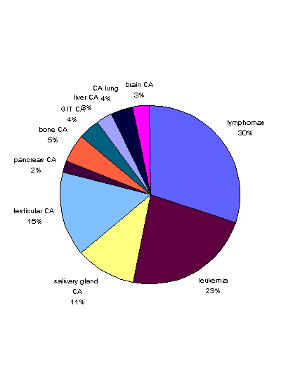
Table (1):- There is relative decline in the percentage
of lymphoma in 1997 in comparison with 1991. It represents about
34.5% in the year 1991, but represents 29.4% in 1997. Also, there
is a decline in the percentage of leukemias and bone cancer in
1997 in comparison with 1991. This may be due to increase in
registration of other types of cancer.
Table (2):- Shows increase in the registration of
different types of cancer cases from the years 1993-1994 to those
participated, but not exposed, which is probably similar to the
pattern in the general population. Also there is a decline in
the percentage of registration in 1997 in comparison to 1991 concerning
lung CA, leukemias and probably this is due to the increase registration
of other cancers such as gastrointestinal cancer, leukemias, lymphomas,
bone CA, and brain CA, which were not registered in 1991. The end result of the exploded weapon could be as dust, fumes, aerosol, or ashes that contain uranium oxide particles. These particles could be transmitted by air or carried with winds and can be airborne for miles before dropping to the ground (Dan Fahey 98).
This can raise the probability of different types
of cancer cases occurrences to those exposed to field explosion
and front units, in tank crews, armored vehicles, air force defense,
artillery, those personnel who were in administrative units, and
in maintenance of destroyed vehicles or evacuation.
Table (2) also shows increase in registration of
cancer cases in those who participated were but not exposed.
This probably can be due to environmental pollution, which might
be similar to the pattern of cases in the general population.
Dust of D.U., as a result of it's explosion, can
fall on the ground and personnel can inhale it or it enters the
body through GIT. In addition, some of the bullets that miss
their targets fall on the ground and could be considered as environmental
pollutant that can contaminate food and surface water sources
and ground water.
Its effect on environment depends on the type of
soil and the degree of solubility of uranium oxide particles.
(Dan Fahey 98).
Table (3):- shows statistically significant odds
ratio of different types of cancer cases to those involved and
exposed to an explosion in the battlefield. From an epidemiological
point of view, it is interpreted that there is a positive association
between the explosion (exposure to D.U.) and cancer cases.
Odds ratio was very high in lymphomas which was 5.6,
followed by 4.8 in cases of leukemias, and 4.5 in brain CA.
In fact, the odds ratios of other cancer cases in
relation to exposure to an explosion (the factor under study)
also have statistically significant values as shown by Table
(3), but in a lesser degree such as in cases of lung, gastrointestinal,
liver, and bone cancers.
The probability of this association as shown from
odds ratio could be related to the causative agent (D.U.).
D.U. contains alpha particles, the insoluble part
of these particles represent about 83.52%. This quantity, once
entered the body of the exposed person, will stay there and consequently
ionize body organs and tissues surrounding it, causing different
types of cancer. (Dan Fahey 98).
The soluble part of alpha particles, which represent
48%, is transmitted to the human body by absorption and through
blood circulation. Only about 60% will be excreted by urine and
stool within a few weeks. The residual 20% will be deposited
in the bones and the other 10% will be distributed to the other
body organs, especially the liver, causing ionization of the tissues
of the organs and disturbance of their function. This, consequently,
may cause different types of cancer cases (Los Alamos National
Laboratories 1996).
The soluble part of alpha particles inside the body
of human being has a chemically toxic effect on DNA of reproductive
cells which causes congenital anomalies and destructive effects
on reproductive mechanisms resulting in primary and secondary
sterility (Dan Fahey 98).
The mechanism of cause and effect needs further laboratory
and experimental studies of those exposed to the causative agent
through examination of its effect on chromosomes.
It is stated by U.S. Army Environmental Institute
Report 1995 and Department of Veterans Affairs 1997, that "D.U.
has an effect on changes of the shape of chromosomes in terms
of increase in sister chromatid exchange" and that was proven
through lymphocyte culture of two groups of people working in
uranium manufacturing in U.S. The end result of the study shows
that the group of workers exposed to soluble type of uranium have
a statistically significant rise in chromosomal aberration and
sister chromatid exchange in comparison with group of workers
exposed to soluble and insoluble type of uranium.
The probability is that D.U. has a chemically toxic
and radiological effect due to the alpha particles it contains:
consequently causing infertility, congenital anomalies, low birth
weight of babies of fathers and mothers exposed to D.U.
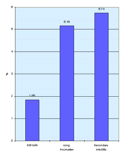
This may explain the 222 cases of congenital anomalies
and stillbirths related to military personnel who were exposed
to the explosions in the southern region of Iraq as shown in Graph
15.
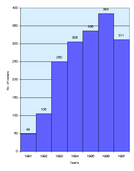
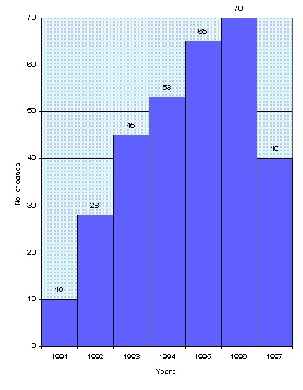
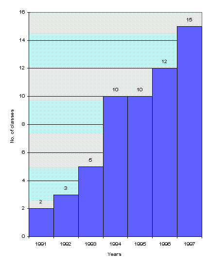
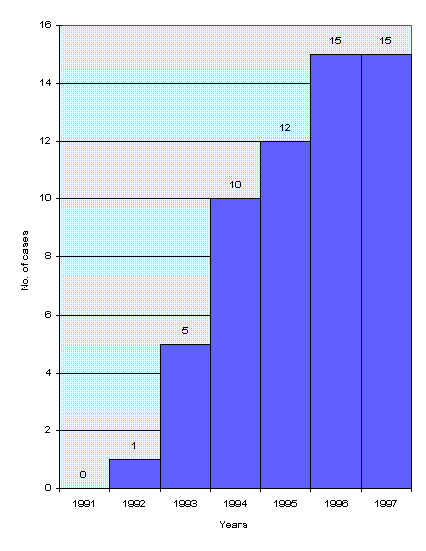
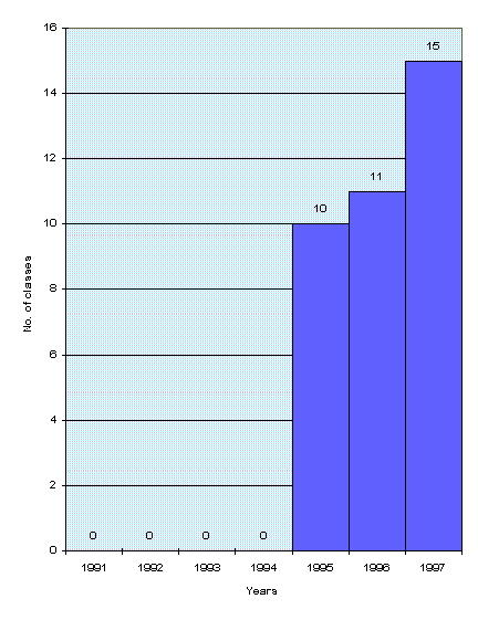
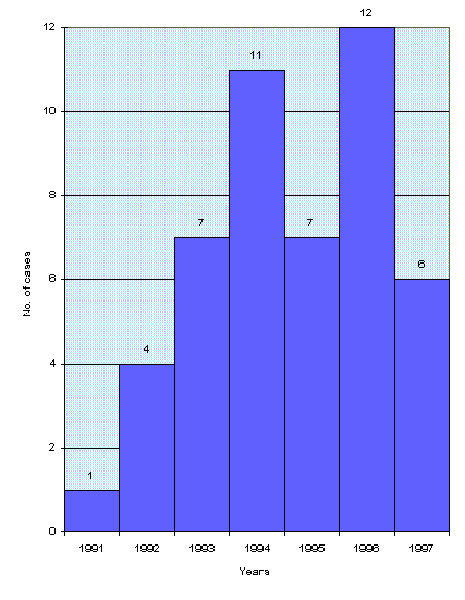
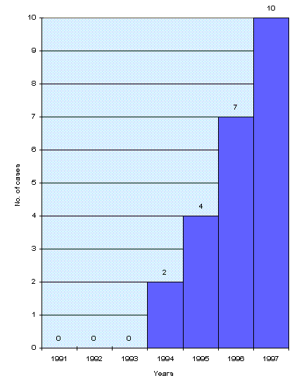
Graph (1):- represents subsequent rise in registration
of total cancer cases in those exposed and not exposed.
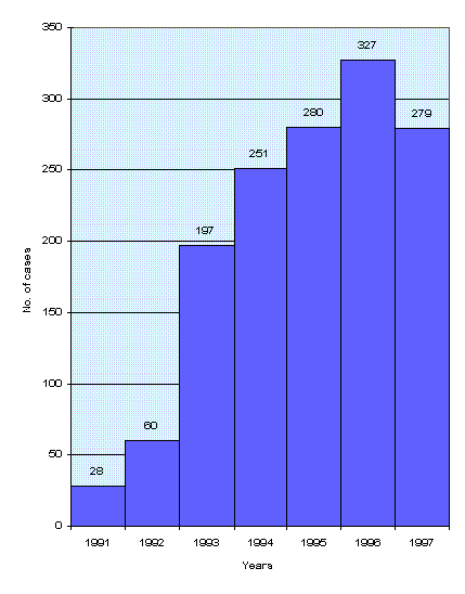
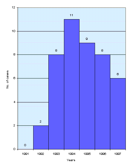
Graph (2):- shows subsequent rise in registration
of different types of cancer cases in those who participated and
were exposed to explosion from the years 1991 up to 1997.
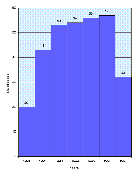
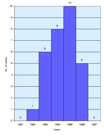
Graph (3):- represents the total number of different
types of cancer cases in military personnel who participated but
not exposed to explosion,. Graph (3) also shows subsequent rise,
but in a lesser degree, probably due to environmental pollution.
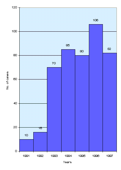
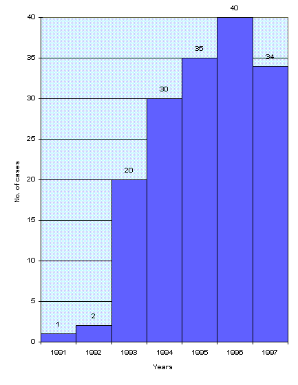
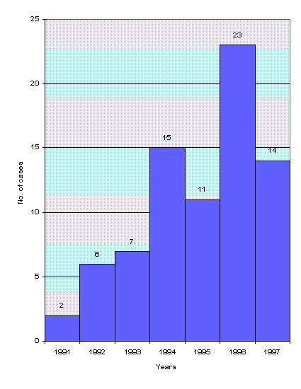
Graphs (4-13):- show an increase in registration
of different types of cancer cases from the year 1992 forward
of those military personnel who were exposed to D.U. explosions
including lymphomas, leukemias, lung CA, brain CA, GIT CA, bone
CA, testicular CA, pancreatic CA, salivary and liver CA. In fact,
the real rise was from the year 1993-1994 forward which shows
the effect of factor under study which needs a lapse of years
to show it's effect in terms of it's ionizing radiation of alpha
particles constituent.
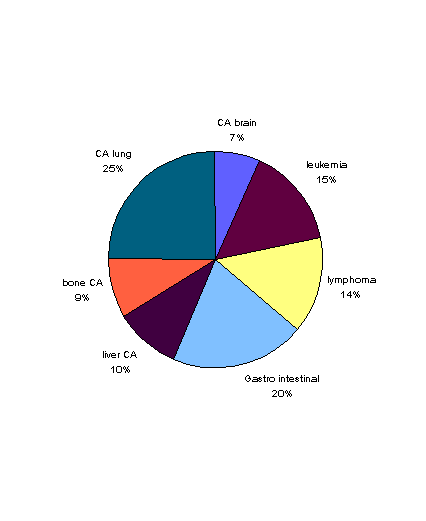
Graph (14):- shows the percentage of different types
of cancer cases in military personnel who participated and were
exposed to the explosion. The most common type registered was
lymphomas, leukemias, lung CA, brain CA respectively. This might
give a clue about the causative agent and its pathogenesis.
Graph (15):- shows the percentage of still birth,
congenital anomalies, and secondary infertility in families of
military participants exposed to D.U. explosions.
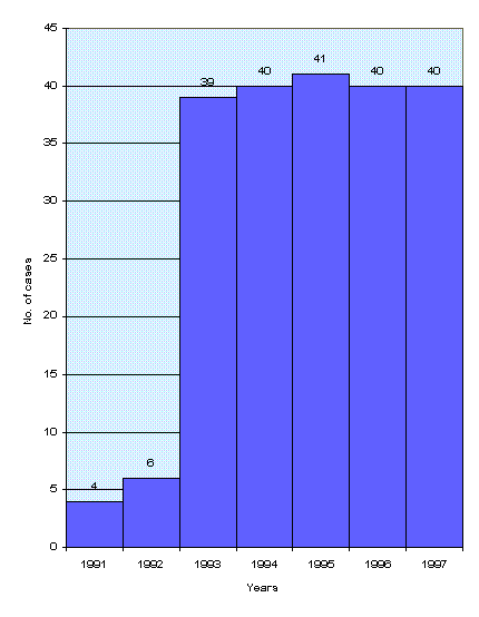
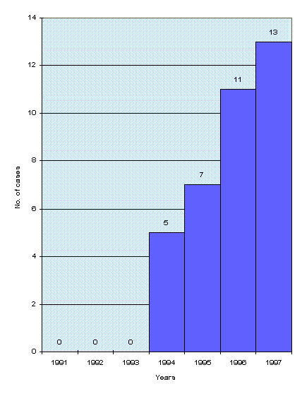
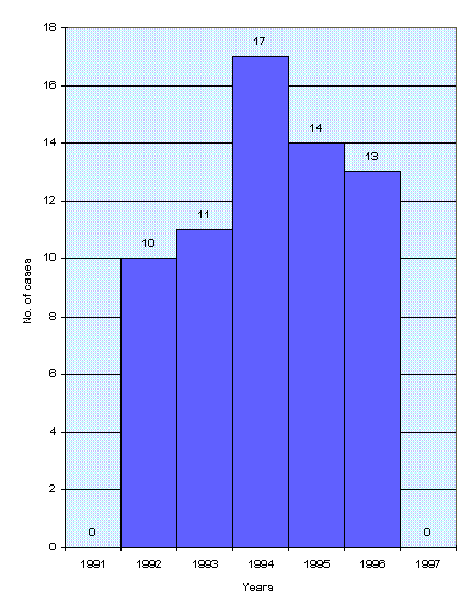
Graphs (16-22):- show the pattern of total number
of different types of cancer cases in military personnel who participated,
but not exposed to D.U. explosion. These graphs also show an
increase in registration of cancer cases among them, but in a
lesser degree, also after a lapse of 3-4 years followed by subsequent
rise.
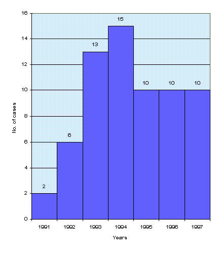
Graph (23):- shows the registration of different
types of cancer cases among unexposed personnel who participated
in the southern region of Iraq during U.S. and British attacks.
The common cancers registered were lung CA, GIT, leukemias and
lymphomas. It may be interpreted in the following manner:
The dosage of exposure due to environmental pollution
may play a role in the pathogenesis of these diseases through
inhalation and ingestion of high doses of particles of the causative
agent.
Further studies are required to isolate the causative
agent a principal cause for such problems, especially to determine
the effect of environmental pollution.
Conclusion:
The quantity of D.U. weapons used by U.S. and British
forces against Iraq in 1991 was estimated to be approximately
5000-6000 rounds of 120 mm and 940,000 bullets of 30 mm. Its
weight was approximately 630,000 pounds (Dan Fahey 98).
The effluent of this weapon is the dust of uranium
oxide resulted in pollution of thousands of targets hit by this
sort of weapon and surrounding areas in the southern region of
Iraq, Kuwait, and Saudi Arabia. The study of tables and graphs shows the change in the pattern of different types of cancer cases in military personnel who participated in the southern region of Iraq (exposed or not exposed to an
explosion). They also show positive odds ratio of
cancer cases in relation to the explosion which indicates the
association between the factor under study (D.U.) and these cases.
From epidemiological criteria of effluent, the usage
of such weapons creates dust or aerosol as a result of explosion
of D.U. weapon in the field, and may be transmitted directly to
the human body through respiratory system or gastrointestinal
system or contaminate wounds directly or through contaminated
shrapnel.
The indirect method of transmission is through pollution
of the environment in which the soluble part of alpha particles
deposited on the soil. In addition, the bullets that missed their
targets and fell on the ground can pollute food, surface water,
and ground water. (U.S. Army Chemical School, D.U. training 1995).
The significant odds ratio of lymphomas, leukemias,
brain CA, which were 5.6, 4.8 and 4.5 respectively in the exposed
personnel.
Proposals: Provision of epidemiological and clinical studies to all health problems related to the use of D.U. weapons The need for a prospective study to compare the exposed personnel to D.U. with group of personnel who were unexposed. Study should be followed for at least 2 years to directly determine the odds ratio. Provision of training survey for doctors and allied personnel for such problems. Studies should concentrate upon high risk groups and should be studied long term. Utilize all the experiences of countries that were exposed to ionizing radiation weapons in the world. Provisions of laboratory studies of chromosomes to confirm the chemically toxic effect of ionizing factors. Clinical epidemiological screening of urinary tract system to those exposed to D.U. to identify the ill effects. Identify the military troops which are at risk, and expose them for epidemiological and clinical studies.
Screening of all Iraqi cities as to the level of
contamination, not only the southern region.
REFERENCES: 1. Dan Fahey, case narrative, depleted uranium exposures, last update March 1998, p. 1-180. 2. Development of depleted uranium training support packages, Tier 1. General Audience, U.S. Army Chemical School, October 1995, B-5 to B-6. 3. Dietz L.A., Estimate of radiation dose from a depleted uranium oxide particle, 1991, reproduced in Appendix, Uranium Battlefields. 4. International Bone Seeking Radio Nuclides and Monocytes Counts, International prospective in public health, Dr. Rosalie Bertell, Vol. 9, 1993, p.21. 5. Kinetic Energy Penetrator Environmental and Health Considerations (Abridged). Science Application, International Corporation, July 1990, Vol. 1, 2-2.
6. Middle East trip provides useful information exchange,
Gulf Link, Jan. 28, 1998. Walid Al-Tawil interview conducted in
Baghdad Iraq, May 1995.
Prof. Mikdam M. Saleh, Ph.D. Ahmed J. Meqwar
Abstract Weapons containing "Depleted Uranium" (DU) were used, for the first time during the "Mother of All Battles" (Operation Desert Storm) by the allied forces against military targets, industrial installations, and civilian populations. The long-term effects of DU on the Iraqi environment, especially in the southern region, were examined and analyzed. The exposure rate to external gamma radiation was measured in six selected regions in the study area (Basra, Zubair, Safwan, Jabal Sanam, and north and south Rumaila), where (154) biological samples (plant and animal tissues), (128) soil and (60) water samples were collected from these regions. Gamma spectrometric analysis of plant samples indicated the presence of some isotopes of the U-238 series in (36.4)% of the total samples, which were collected from the study area. Some of the wild plant samples (Haloxylon salicornicum and Stippa capensis) tended to have high concentrations of Th-234 and Ra-226 (with several samples whose concentrations exceeded those taken from regions with natural background level). The minimum concentrations of Th-234 and Ra-226 were (28, 62) Bq.Kg-1, respectively, were found in the Haloxylon salicornicum samples, and the maximum (249, 275) Bq.Kg-1, were found in the Stippa capensis samples. Other samples concentrated Bi-214 and Pb-214, with concentration of (1-3) times the natural background level. The "Transfer Coefficient" (TC) between soil and plant for U-238 and Ra-226 were measured, for the first time in Iraq (as far as can be ascertained). TC values for U-238 and Ra-226 were [0.17(54.2%), 0.37(79.6%)], respectively, and are in good agreement with corresponding values reported in the literature. The internal and external doses that were delivered to the population in the study area have been calculated. The average doses over the period from (1991-1996) due to inhalation, ingestion of meat and milk, and external exposure, were (87.4, 0.25, 0.20, 0.13) mSv, respectively in each of Basra and Zubair, (173.5, 0.51, 0.39, 0.27) mSv, respectively, in Safwan. The result of these calculations showed that the dose delivered to infants and children (less than 15 years old) represent (70)% of the total dose delivered to the general population. This study, also, predicted the fatality rate for the newly born (300) and for the general population (800) due to leukaemia, over the period from (1991-1996). The expected incidence (after 10 years latent period) for lung cancer was estimated at (44)% of the total population, while for fatal or non-fatal cancer and hereditary damages at (5)%. The "Risk Coefficient" (RC) of mortality due to leukemia, from 1991-1994, was estimated at (2.67 Sv-1). This is in agreement with many values published in the literature. Calculations showed that an estimated (845,100) tons of edible wild plant were contaminated with radioactive materials (U-238 series), and (31)% of the animal resources were exposed to internal radioactive contaminants that exceeds the natural background level. An estimated area of (1044800)m2 in which the soil organisms were exposed to radioactive contaminants, which exceeds the threshold level for damage. Lastly, this study estimated the total damages incurred to the population and biosphere in the study area, at the sum of (372.2) billion dollars.
The effect of the war of the American and the
affiliated forces against Iraq on the distribution and elevation
of cancer diseases in Mosul. Mohemid Maddadah Al-Jebouri, M.Sc. Ph.D. M.I. Biol.
Department of Microbiology, College of Medicine,
University of Tikrit, Tikrit Iraq Ibraheem Abdul-Kareem Al-Ani, B.Sc., M.Sc.
Department of Biology, College of Science, University
of Al-Anbar, Ramadi, Iraq Salih Abed Al-Joumaily, M.B., Ch.B., Ph.D.
Department of Pediatrics, College of Medicine, University
of Mosul, Mosul, Iraq
All correspondence to: Prof. M.M. Al-Jebouri, Department of Microbiology, College of Medicine,
University of Tikrit, Tikrit, Iraq
Abstract:
A total of 1094 cancer cases were reported in four
hospitals and/or medical centers of Mosul city during 1974-1990
and 1997-1998. The present study showed that males were under
risk of cancer more than females. Deep x-ray as a treatment tool
for tumour was the most common. The frequency of incidence of
cancer diseases elevated 5-fold after the war in 1991. Lung,
leukemia, breast, skin, lymphoma and liver cancers elevated after
the war. This elevation could be due to the depleted uranium
weapons used by the allied forces against Iraqi troops and citizens.
Other environmental contaminants with health hazards should be
not excluded.
Introduction:
Radiation can cause cancer. Highly energetic ionizing
radiation (capable of knocking electrons out of atoms and molecules)
is the most hazardous. But non-ionizing radiation such as ultra-violet
light, plays an important role in the genesis of human skin cancer.
Survivors from Hiroshima and Nagasaki showed a marked elevation
in the incidence of leukemia five years after the explosions in
1945. After a later period, the incidence of breast, thyroid,
lung, and bowel cancers were found to be modestly elevated. (Currie
and Currie, 1982) The effect of radiation on man can be observed
as weakness, age shortening, redness of the skin, and the elevation
of incidence with leukemia (Raymon, 1987). The effect of radiation
on human cells appears within 10-16 second, usually
called the direct physical effect of radiation. The biological
effect of radiation not only depends on the dose, but also depends
on the relative biological effect (R B E) which is consequently
dependent on the type of radiation used. This is defined as the
quality factor. (Raymon, 1987. Krane, 1987). The present study
is an assessment of the effect of the American and affiliated
forces attacks using the depleted uranium munitions and the rate
of incidence of cancer diseases among the Iraqi peoples by comparing
the incidence of cancers before and after the war in 1991.
Patients and Methods:
The cancer diseases were recorded in four hospitals
and/ or medical centers for cancer treatment and management in
Mosul city from August 1989 to March 1990. The same work was
repeated in the same hospitals and medical centers during August
1997 to March 1998. Type of cancer, sex of patients, and type
of therapy was studied. The two most prevalent types of cancer
diseases were also examined and statistically analyzed (Al-Ani,
1990).
Results: Table (1) shows that the rate of incidence of cancer was increased almost 3-fold after the attacks of the American and affiliated forces during 1991. Males were under risk of developing cancers more than females before and after the war. Table (2) shows that most of the cancer patients were treated with deep x-ray. Cytotoxic drugs were also used for treatment of cancer patients either alone or in combination with deep x-ray. Table (3) shows the distribution of various cancer diseases among the patients studied before and after the war. The lung cancer was prevalent with respect to other cancers, and the incidence frequency among males before and after was 20.5 and 25.7 percent, respectively. Lymphoma, leukemia, breast, and larynx cancers were also prevalent with different rate of incidence among males and females before and after the aggression. Table (4) shows the prevalent cancer disease among males and females before and after the war. The present study shows that skin cancer was one of the common diseases disseminated among people, particularly males. This disease was more prevalent after the aggression against Iraq. Not one case was reported among females before the war. The incidence frequency of skin cancer among females after the war was almost 9%. The incidence of breast cancer was also elevated after the war.
Table (5) lists the cancer diseases which are elevated
after the war against Iraq. The lung cancer was elevated among
males from 20.5% to 25.7%. There was no lung cancer cases reported
among females from before the war, but the frequency of incidence
increased up to 3.6% of the total cancer diseases reported after
the war. The cancer of larynx, skin, and liver among females
was reported after the war only. Breast cancer was also elevated
from 21.8% to 27% after the war. Leukemia became prevalent (9%)
among males after the attacks of the affiliated forces. Thyroid
cancer was also elevated from 0.8% to 2% of the total cancer cases
reported in Mosul hospitals after the war.
Discussion:
The present study showed that males were under risk
of developing cancer diseases more than females, and the statistical
differences were significant (P< 0.05). The same conclusion
was reported by Ibraheem and Majeed (1987), Wilson and Patterson
(1989) and Al-Irhayim and Saleem (1990). The explanation for
such difference might be due to occupational, environmental, anatomical,
and physiological differences between males and females (Doll,
1977). Moreover, the elderly peoples were under risk of cancer
more than other age groups. This could be due to the physiological
and immunological change at this age. Furthermore, the present
study revealed that solid tumours were more prevalent, and the
same results were concluded by Elting (1986). However, the distribution
of the cancer diseases among males and females before and after
the war was different. This difference could be due to the change
of environmental conditions and contamination by declared and
undeclared weapons used by the allied forces against Iraq, e.g.
the depleted uranium weapons. The weapons used against the republican
guard forces produced ionizing radiation leading to possible
biological effects on the Iraqis (Al-Jomialy 1996). In 1996,
Al-Jomialy found a remarkable increment in the uranium concentration
especially in Al-Muthanna and the Thee-Qar provinces where the
republican guard forces were concentrated. However, lung, larynx,
lymphoma, bladder, skin, stomach, breast, uterus, leukemia, thyroid,
and liver cancers were all showing and the incidence of the diseases
after the war were elevated.
References: 1. Al-Ani, I.A.K. (1990). Bacteriological studies on cancer patients in Mosul district. M.Sc. thesis, University of Mosul. 2. Al-Jomialy, F.M.A. (1996). Detection of depleted uranium in soils from UM - L - Maalrik battlefield by using CR.-39 detector. M.Sc. thesis. University of Mosul. 3. Al-Irhayim, B. and Saleem, S.H. (1990). Cancer in first two decades of life excluding leukemias - A pathological study of 300 cases in Mosul. 232-237. 4. Currie, G. and Currie, A (1982). Cancer - The Biology of Malignant Diseases. Edward Arnold. London. 5. Doll, R. (1977). Introduction. In II.II.Illatt. J.D. Watson and J.A. Winsten (ed s). Origin of human lab. Cold Spring Harbor, New York, pp. 309-330. 6. Elting, L.S.; Body, G.P.; Fainstein, V. (1986). Polymicrobial septicaemia in the cancer patient. Medicine, 65: 218-255. 7. Ibraheem, K.S. and Majeed, A.A. (1978). Cancer in the north part of Iraq (Ninerveh Province). Iraqi Medical journal, 35:63-66. 8. K.S. (1987). Introduction - Nuclear Physics. John & Sons, Inc. London. 9. Raymon, M. (1987). Nuclear Energy. second edition, Pergamon Press, London. 10. Wilson, R. and Patterson, B.A. (1989). Pattern of malignant tumours in King Fahad Hospital, Al-Baha, Saudi Arabia, Saudi Medical Journal, 10: 498-502.
Table 1. Distribution of cancer patients in the
Mosul Hospital.
Table 2. Distribution of cancer patients according to the
type of therapy.
Table 3. Distribution of different cancer diseases among patients in Mosul hospitals,
before and after the War 1991.
Table 4. The distribution of the prevalent cancer diseases
before and after the war in 1991
Table 5. The cancer diseases with increased incidence after
the war in 1991.
This information is copied and as such is a mirror site from : http://asterix.phys.unm.edu:8000/dureport/dureport.html Also see for lots of info : http://asterix.phys.unm.edu:8000/
| |||||||||||||||||||||||||||||||||||||||||||||||||||||||||||||||||||||||||||||||||||||||||||||||||||||||||||||||||||||||||||||||||||||||||||||||||||||||||||||||||||||||||||||||||||||||||||||||||||||||||||||||||||||||||||||||||||||||||||||||||||||||||||||||||||||||||||||||||||||||||||||||||||||||||||||||||||||||||||||||||||||||||||||||||||||||||||||||||||||||||||||||||||||||||||||||||||||||||||||||||||||||||||||||||||||||||||||||||||||||||||||||||||||||||||||||||||||||||||||||||||||||||||||||||||||||||||||||||||||||||||||||||||||||||||||||||||||||||||||||||||||||||||||||||||||||||||||||||||||||||||||||||||||||||||||||||||||||||||||||||||||||||||||||||||||||||||||||||||||||||||||||||||||||||||||||||||||||||||||||||||||||||||||||||||||||||||||||||||||||||||||||||||||||||||||||||||||||||||||||||||||||||||||||||||||||||||||||||||||||||||||||||||||||||||||||||||||||||||||||||||||||||||||||||||||||||||||||||||||||||||||||||||||||||||||||||||||||||||||||||||||||||||||||||||||||||||||||||||||||